Intrinsic optical imaging
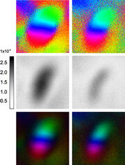
We are using intrinsic optical signals (IOS) in vivo to analyze brain activity following sensory stimulation. Therefore, reflected light from the brain surface is registered by a video camera. Areas with increased activity appear darker compared to their surrounding (see figure). The signal is based on the strong coupling between neuronal activity and blood flow, the so-called neuro-vascular coupling.


Fig.: Left: schematic drawing of the experimental setup. The brain surface is illuminated by a light source and the reflected light is detected by a CCD-camera. Right: Intrinsic optical signals following sensory stimulation of the contralateral eye (left column) and ipsilateral eye (right column). Upper panels reflect phase maps, middle panels represent activity maps and lower panels are the superposition of both.
References:
- Weiler S, Rahmati V, Isstas M, Wutke J, Stark AW, Franke C, Graf J, Geis C, Witte OW, Hübener M, Bolz J, Margrie TW, Holthoff K, Teichert M (2024) A primary sensory cortical interareal feedforward inhibitory circuit for tacto-visual integration. Nat Commun 15, 3081. https://doi.org/10.1038/s41467-024-47459-2

