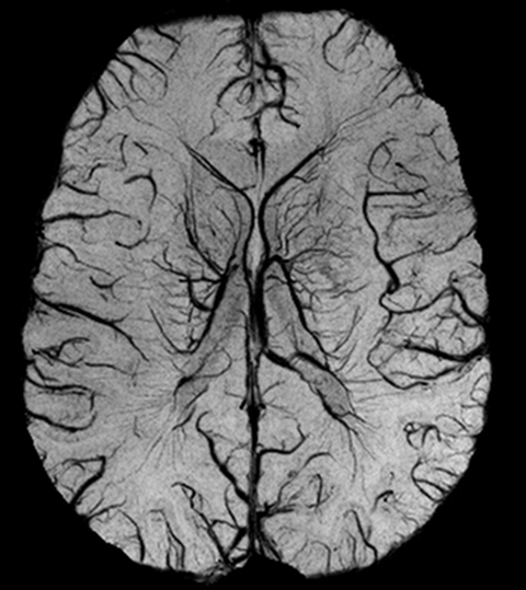
Haacke, E.M., Reichenbach, J.R., Xu, Y., 2011. MRI Susceptibility Weighted Imaging: Basic Concepts and Clinical Applications. John Wiley & Sons.
Haacke, E.M., Xu, Y., Cheng, Y.C., Reichenbach, J.R., 2004. Susceptibility weighted imaging (SWI). Magnetic Resonance in Medicine 52, 612-618.
Reichenbach, J.R., Haacke, E.M., 2001. High-resolution BOLD venographic imaging: a window into brain function. NMR Biomed 14, 453-467.
Reichenbach, J.R., Venkatesan, R., Schillinger, D.J., Kido, D.K., Haacke, E.M., 1997. Small vessels in the human brain: MR venography with deoxyhemoglobin as an intrinsic contrast agent. Radiology 204, 272-277.
Deistung, A., Dittrich, E., Sedlacik, J., Rauscher, A., Reichenbach, J.R., 2009. ToF-SWI: simultaneous time of flight and fully flow compensated susceptibility weighted imaging. J Magn Reson Imaging 29, 1478-1484.
Deistung, A., Mentzel, H.J., Rauscher, A., Witoszynskyj, S., Kaiser, W.A., Reichenbach, J.R., 2006. Demonstration of paramagnetic and diamagnetic cerebral lesions by using susceptibility weighted phase imaging (SWI). Z Med Phys 16, 261-267.
Deistung, A., Rauscher, A., Sedlacik, J., Stadler, J., Witoszynskyj, S., Reichenbach, J.R., 2008a. Susceptibility weighted imaging at ultra high magnetic field strengths: theoretical considerations and experimental results. Magnetic Resonance in Medicine 60, 1155-1168.
Deistung, A., Rauscher, A., Sedlacik, J., Witoszynskyj, S., Reichenbach, J.R., 2008b. Informatics in Radiology: GUIBOLD: a graphical user interface for image reconstruction and data analysis in susceptibility-weighted MR imaging. Radiographics 28, 639-651.
Haacke, E.M., Reichenbach, J.R., Xu, Y., 2011. MRI Susceptibility Weighted Imaging: Basic Concepts and Clinical Applications. John Wiley & Sons.
Haacke, E.M., Xu, Y., Cheng, Y.C., Reichenbach, J.R., 2004. Susceptibility weighted imaging (SWI). Magnetic Resonance in Medicine 52, 612-618.
Rauscher, A., 2005. Phase Information in Magnetic Resonance Imaging.
Rauscher, A., Barth, M., Herrmann, K.H., Witoszynskyj, S., Deistung, A., Reichenbach, J.R., 2008. Improved elimination of phase effects from background field inhomogeneities for susceptibility weighted imaging at high magnetic field strengths. Magn Reson Imaging 26, 1145-1151.
Rauscher, A., Barth, M., Reichenbach, J.R., Stollberger, R., Moser, E., 2003. Automated unwrapping of MR phase images applied to BOLD MR-venography at 3 Tesla. J Magn Reson Imaging 18, 175-180.
Rauscher, A., Sedlacik, J., Barth, M., Haacke, E.M., Reichenbach, J.R., 2005. Nonnvasive assessment of vascular architecture and function during modulated blood oxygenation using susceptibility weighted magnetic resonance imaging. Magnetic Resonance in Medicine 54, 87-95.
Rauscher, A., Sedlacik, J., Deistung, A., Mentzel, H.J., Reichenbach, J.R., 2006. Susceptibility weighted imaging: data acquisition, image reconstruction and clinical applications. Z Med Phys 16, 240-250.
Reichenbach, J.R., Haacke, E.M., 2001. High-resolution BOLD venographic imaging: a window into brain function. NMR Biomed 14, 453-467.
Reichenbach, J.R., Venkatesan, R., Schillinger, D.J., Kido, D.K., Haacke, E.M., 1997. Small vessels in the human brain: MR venography with deoxyhemoglobin as an intrinsic contrast agent. Radiology 204, 272-277.
Sedlacik, J., Helm, K., Rauscher, A., Stadler, J., Mentzel, H.J., Reichenbach, J.R., 2008a. Investigations on the effect of caffeine on cerebral venous vessel contrast by using susceptibility-weighted imaging (SWI) at 1.5, 3 and 7 T. Neuroimage 40, 11-18.
Sedlacik, J., Kutschbach, C., Rauscher, A., Deistung, A., Reichenbach, J.R., 2008b. Investigation of the influence of carbon dioxide concentrations on cerebral physiology by susceptibility-weighted magnetic resonance imaging (SWI). Neuroimage 43, 36-43.
Sedlacik, J., Rauscher, A., Reichenbach, J.R., 2007. Obtaining blood oxygenation levels from MR signal behavior in the presence of single venous vessels. Magnetic Resonance in Medicine 58, 1035-1044.
Sedlacik, J., Rauscher, A., Reichenbach, J.R., 2009. Quantification of modulated blood oxygenation levels in single cerebral veins by investigating their MR signal decay. Z Med Phys 19, 48-57.
Witoszynskyj, S., Rauscher, A., Reichenbach, J.R., Barth, M., 2009. Phase unwrapping of MR images using Phi UN--a fast and robust region growing algorithm. Med Image Anal 13, 257-268.


