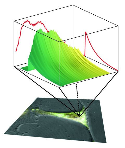
In conventional fluorescence microscopy only the information conferred by the fluorescence intensity is used to delineate microscopic structures or to perform quantitative measurements by the use of fluorescent indicator dyes. However, a fluorophore is not only characterized by the intensity of the emitted light, but also by its absorption and emission spectra, by its lifetime in the excited state and the polarization of the emitted light.
The fluorescence lifetime does not only provide another dimension of contrast, but can also be exploited for functional imaging. The fluorescence lifetime is sensitive to the physical and chemical environment of the fluorophore. As such it is an excellent reporter of its surroundings.
The Biomolecular Photonics Group has established several spectrally resolved fluorescence lifetime imaging techniques based on time-correlated single photon counting (TCSPC) and a streak camera system. Instead of a single fluorescence decay curve, which is obtained by conventional techniques, a fluorescence decay surface can be reconstructed from the data obtained by these techniques (Fig. 1).


